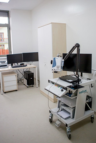
Electroencephalography and electromyography (EEG-EMG)
The EEG-EMG Laboratory includes the following devices:
- 32-channel electroencephalograph (NicOne® V 32 workstation, Natus Medical Inc., Nicolet)
- system for digital polysomnography (incorporating the 32-channel EEG);
- 3-channel electromyograph (Viking EDX®, Natus Medical Inc., Nicolet).
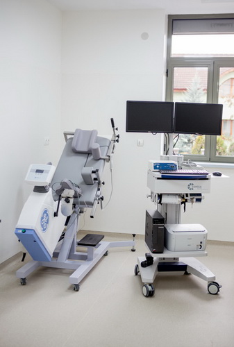
Syncope and other autonomic disorders (SINC)
The Laboratory includes the following devices:
- multifunctional polyphysiograph with multiple recording features (Task Force Monitor 3040i®, GE Healthcare)
- state-of-the-art tilt table, with electrically adjustable height and tilt angle (0° to 85°) (Tilt 2900-00®, GE Healthcare).
The system ensures:
- continuous, real-time, high-resolution ECG recording;
- non-invasive, continuous, beat-to-beat measurement of the systolic, diastolic, and mean blood pressure;
- continuous, beat-to-beat measurement of the heart rate;
- non-invasive, continuous cardiac output and stroke volume measurement using impedance cardiography;
- systemic vascular resistance evaluation;
- autonomic nervous system assessment using baroreflex and heart rate variability.
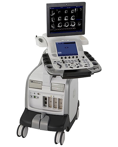
Cardiac and vascular imaging (ECHO)
The Laboratory includes an advanced echocardiograph unique in Romania at this level of concomitant facilities (Vivid E9 BT12®, GE Healthcare), conceived for bidimensional transthoracic and transesophageal adult, pediatric, and neonatal cardiac examination. The device is also suitable for vascular and transcranial examinations, as well as for contrast and interventional echography.
The echocardiograph disposes of the XD Clear technology, which offers high image resolution and allows highly accurate measurements, as well as of a set of softwares for advanced quantifications.
The device is equipped with:
- one matrix transducer (M5S-D), build using the pure wave technology, designed for cardiac adult, pediatric and fetal echography, transcranial examination, stress echocardiography, contrast echocardiography with left ventricular contrast, renal echography;
- one linear transducer (ML6-15-D), designed for vascular, breast, soft tissues, musculoskeletal, thyroid, scrotal, and rodent echography;
- one convex transducer (4C-D), designed for abdominal, obstetrics-gynecology, urology, vascular, fetal cardiac, pelvis, and renal echography.
Standard examination modes include:
- 2D mode, with automatic tissue optimization, which can be manually implemented by pushing a single button; automatic optimization can also be activated dynamically by selecting he CTO (continuous tissue optimization) option;
- M-mode, anatomic M-mode, curved anatomic M-mode, color M-mode, and color anatomic M-mode;
- pulsed wave/HPRF, continuous wave, and color Doppler;
- spectral tissue and color tissue Doppler;
- myocardial deformation quantification by speckle tracking (AFI – Automatic Function Imaging – option); the results can be presented in the “bullseye” manner;
- contrast echography (LVO contrast option);
- Q analysis;
- B-flow for vascular imaging.
The echocardiograph:
- disposes of Z-scores for pediatric examinations;
- can be updated for stress echocardiography;
- can be updated for 3D echocardiography;
- is compatible with automatic evaluation of the intima-media thickness software.
The external working station includes all post-processing, measurement, and analysis tools available on the echocardiograph.
Electrocardiography (ECG)
The ECG Laboratory includes the following devices:
- 12-lead electrocardiograph (MAC 5500®, GE Healthcare) offering the possibility of using 3 additional leads.
- The device is highly sensitive in recognizing paced rhythms, and disposes of a number of specialized softwares – acute coronary syndrome prediction, QT-interval analysis.
- The device is also suitable for performing signal-averaged ECG, and disposes of a dedicated software for measuring late ventricular potentials and for signal-averaged P-wave analysis.
- The device can also perform vectorcardiographic analysis, disposing of a dedicated software for vectorcardiographic signals (P, QRS, ST-T vectors/loops).
- Five 12-lead Holter ECG monitoring systems (CardioMem 3000®, GETEMED/GE Healthcare).
- The devices allow up to 48-hours of continuous 12-lead ECG monitoring.
- The software (CardioDay®, GETEMED/GE Healthcare) is designed to:
- classify and edit QRS events, and rank the events based on their severity;
- detect paced rhythms;
- recognize atrial fibrillation;
- perform complex ST-segment analysis;
- perform complex, beat-to-beat QT-interval analysis;
- perform heart rate variability analysis in both time and frequency domains;
- perform T wave alternans analysis;
- perform heart rate turbulence analysis;
- perform deceleration capacity assessment.
Stress testing (STRESS)
The STRESS Laboratory includes the following devices:
- bad-like stationary bicycle for pedaling in semi-sitting position, with adjustable tilting, adapted for concomitant echocardiographic examination (eBike EL®, GE Healthcare);
- dedicated system for the recording and interpretation (CardioSoft®, GE Healthcare) of effort electrocardiogram;
- ergospirometry system (Cortex Metalyzer 3B®, GE Healthcare) and analysis software (MetaSoft®, GE Healthcare);
- automatic biphasic external defibrillator (Responder 2000®, Cardiac Science).
The system ensures automatic blood pressure measurement.
The system performs:
- automatic and manual QRS detection and analysis;
- automatic detection of cardiac arrhythmias;
- complex ST-segment analysis – ST-segment amplitude, slope, integral, index, ST/HR slope, ST/HR loops, ST/HR index;
- T wave alternans analysis during stress testing;
- vectorcardiographic analysis.
The system also allows:
- post-testing E, J, and post-J points selection and analysis;
- cardiopulmonary analysis during stress testing;
- VO2max, FIO2, FEO2, METS, AT, RR, VE/VO2, and PETO2 measurement.
The system includes a software dedicated to ergospirometic analysis in athletes.
Metabolic disease testing (METAB-DZ)
The METAB-DZ Laboratory includes the following devices:
- two- and four-syringe pumps for highly accurate and precise administration of intravenous fluids (PHT Ultra®, Harvard Apparatus);
- volumetric infusion systems for intravenous fluids (Infusomat fmS®, B Braun Melsungen AG);
- point-of-care testing system for glucose, lactate, and triglycerides from capillary blood (Accutrend Plus®, Roche Doagnostics GmbH);
- mechanic body weight and height meter;
- continuous interstitial glucose monitoring systems (iProTM2®, Medtronic MiniMed).
The digital recorders of the continuous interstitial glucose monitoring systems collect and store the data received from implanted glucose sensors. Designed specifically for research purposes, the systems have high accuracy. The recorded data is not available for the patient to see, but the stored information can only be accessed at the end of the recording interval. The system is able to synchronize the data with those obtained using conventional glucometers. The implanted sensors allow continuous glucose measurement for up to 7 days.
Stationary
The stationary disposes of four 2-bed wards, designed for short-term (up to 24 hours) monitoring of patients included in various studies.
Each bed is equipped with a vital signs (heart rate, non-invasive blood pressure, oxygen saturation) monitor (B 20®, GE Healthcare), connected to a central monitoring station (C-XPNET 7®; N-REC2®, GE Healthcare).